
Digitizing biological specimens has long been constrained by throughput — a single micro-CT scan can consume an entire working day per sample. A team of researchers has now collapsed that timeline by orders of magnitude, combining a synchrotron particle accelerator, robotics, X-ray imaging, and AI to produce detailed 3D models of 800 ant species in one week.
The project, published in Nature Methods on March 5, 2026, was led by Evan Economo of the University of Maryland’s Department of Entomology and Thomas van de Kamp at the Karlsruhe Institute of Technology (KIT) in Germany. The collaboration drew ant specimens from museums, partner institutions, and specialists worldwide, preserved in ethanol and organized by species and caste before transport to KIT for imaging.
At the facility, a synchrotron accelerator generated an intense X-ray beam while a robotic sample changer rotated each specimen and swapped in the next every 30 seconds. That workflow produced stacks of 2D images later assembled into complete 3D models. According to the announcement, the team scanned 2,000 specimens in a single week — a pace that would have required six years of continuous operation using a conventional lab-based CT scanner, according to first author Julian Katzke, a graduate of Economo’s lab at the Okinawa Institute of Science and Technology (OIST) in Japan.
From Raw Scans to Lifelike Models
Speed introduced its own problem. Specimens arrived in twisted or awkward positions that produced distorted poses in initial scans — far from the natural stances researchers wanted for a usable reference library. To correct this, students enrolled in James Purtilo‘s software engineering course at UMD developed AI tools to automate pose estimation, adjusting scanned images so ants appear as they would in the field.
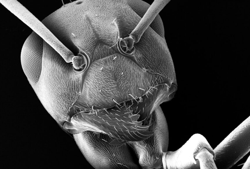
The resulting models expose anatomy that was previously difficult to study at this scale: muscles, nervous systems, gastrointestinal tracts, and stinger apparatus are rendered at high resolution, with portions of the exoskeleton digitally removable for interior inspection. The raw data is publicly available for download, and an integrated viewer lets users explore completed models online under the project’s name, Antscan.
Implications Beyond Entomology
Economo frames the work as a methodological proof of concept rather than a purely entomological exercise. “The value of this study is not only about ants — it’s much broader,” he said. “When specimens are digitized, we can build libraries of organisms that can streamline their use from scientific laboratories to classrooms to Hollywood studios.”

The team’s prior reliance on micro-CT scanning stretched back more than a decade, with individual scans taking up to 10 hours per specimen. The synchrotron-based pipeline compresses that investment by a factor that makes large-scale biodiversity digitization operationally feasible for the first time. The researchers say the Antscan framework could serve as a template for similar high-throughput digitization efforts across other taxonomic groups.
Photo by Mathew Schwartz on Unsplash
This article is a curated summary based on third-party sources. Source: Read the original article